All the images in this brick are in T1
Thalamus
The thalamus is a grey matter structure, and therefore appears darker than surrounding white matter on T1.
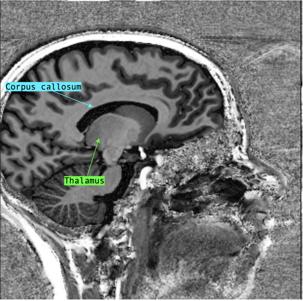
Sagittal
The thalami are large, oval grey matter structures best seen slightly off-center from the midline, immediately lateral to the third ventricle. It is found immediately underneath the fornix, a white matter and therefore paler structure that curls like an arch above the thalamus and framing the lateral ventricle with the corpus callosum. Inferior to the thalami are the midbrain and the rest of the brainstem.
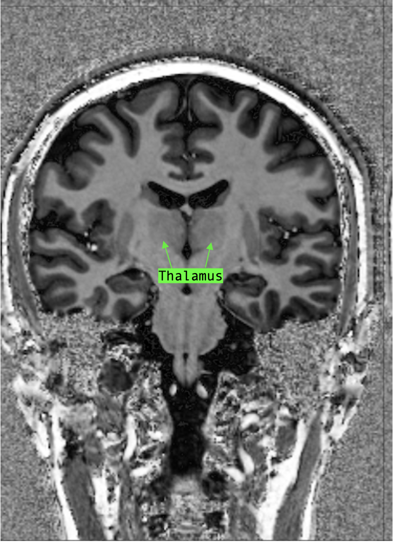
Coronal
The thalami are seen very well on coronal section. They generally come into view at a level when the third ventricle is visible and continue to be visible until the trigone (of the lateral ventricles) come into view posteriorly. The thalami bilaterally sandwich the third ventricle and are the floor of the lateral ventricles. They are delimited laterally by the internal capsule, a white matter structure, which permits some visual contrast to distinguish the thalami.
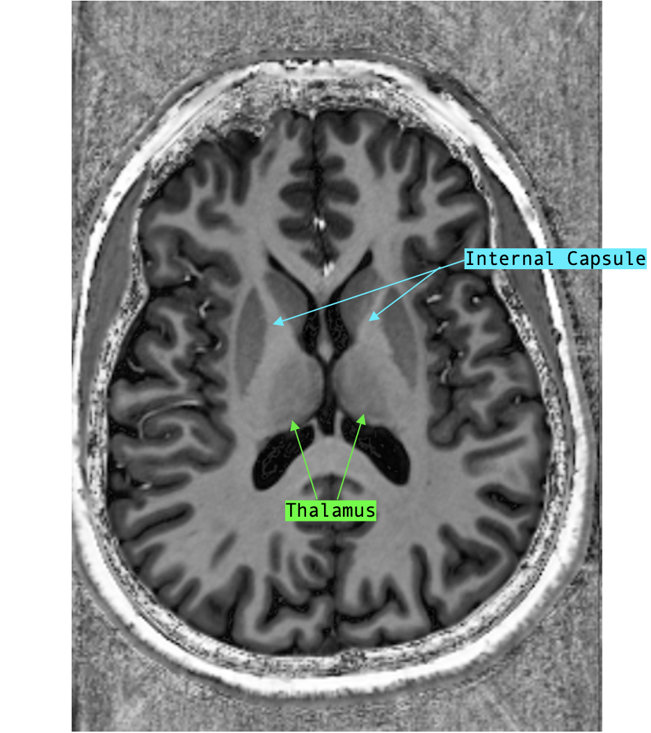
Axial
To find the level where the thalami are, start from the top of the brain (most superficial section) and scroll inferiorly. The lateral ventricles will come into view and they will first appear continuous. The thalami come into view when the anterior horn and posterior horn of the lateral ventricles “split”. Once again, the thalami are on each side of the third ventricle and are delimited laterally by the X-shaped internal capsule. They are in between the anterior horn and posterior horn of the lateral ventricles and appear “rounded in” towards the midline.