The basal ganglia are grey-matter structures surrounded by the deep white matter of the cerebrum. Therefore, on T1, they will appear darker than the surrounding white matter tracts.
*All MRI images in this brick are in T1*
Axial
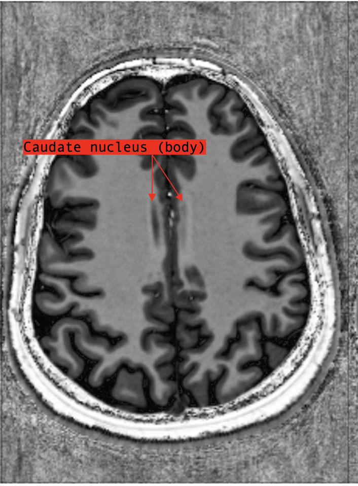
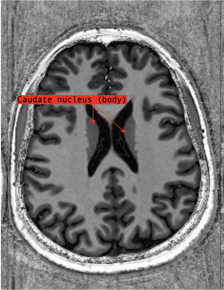
Going from superior sections towards inferior, we first encounter the body of the caudate which appears as a sliver of grey matter nestled into the convexity of the central part of the lateral ventricles.
Remember, the caudate nucleus (see Basal Ganglia) is a 3D “C”-shaped structure that follows the curvature of the lateral ventricles.
Moving inferiorly, the sliver of grey matter gets bigger, moves more anteriorly and gradually appear to jut into the anterior horns of the lateral ventricles: at this point, we are observing the head of the caudate.
Around the levels where the body of the caudate transition into the head of the caudate, the lenticular nuclei (see Basal Ganglia) become visible.
Roughly diamond-shaped, the lenticular nuclei are separated from the caudate nucleus and the thalamus by white matter tracts called the internal capsule that, when looked at bilaterally, form an “X”-shape.
Laterally, the lenticular nuclei are separated from the grey matter of the insular cortex by another set of white matter tracts called the external capsule.
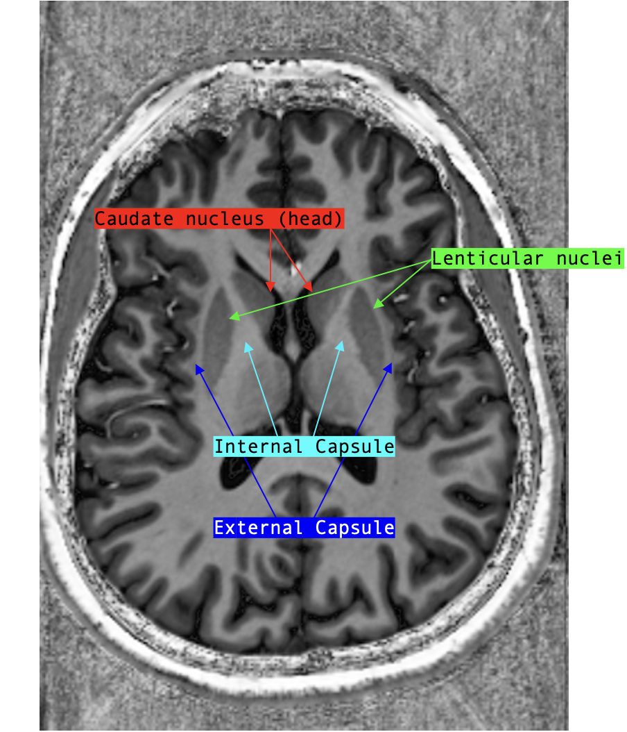
The internal and external capsules are really useful landmarks to look for, as they literally delineate the lenticular nuclei.
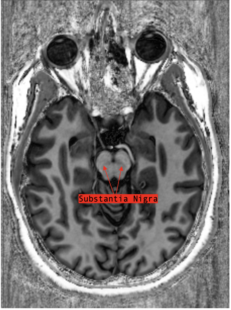
Further inferior, we reach the midbrain.
At the levels where both the midbrain and third ventricle are visible, we can find the substantia nigra.
If you think of the midbrain like a Mickey Mouse head, then the substantia nigra are the dark lines transecting the bases of both ears.
Coronal
Moving anterior to posterior, the first basal ganglia structure we encounter is the head of the caudate.
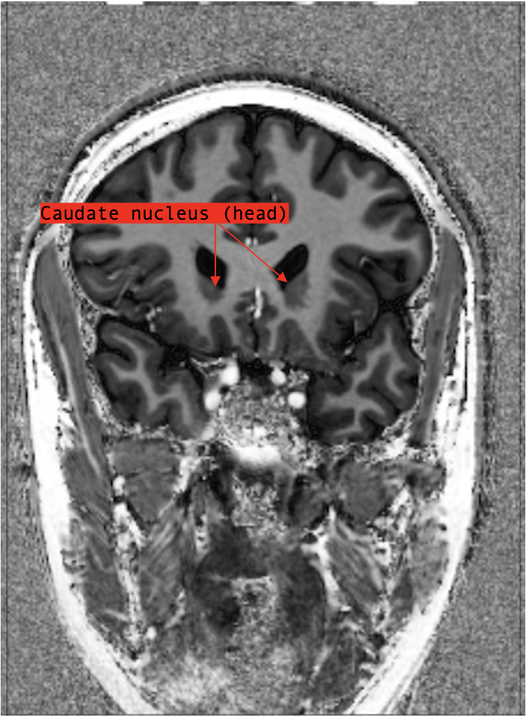
The caudate nucleus is largest anteriorly and gradually tapers posteriorly.
And so, in these initial sections, the head of the caudate appears as a very large grey matter structure pushing up on the floors of the lateral ventricles.
As the sections move more posterior, the head of the caudate seems to get “split” by a diagonal white matter tract (no other than the internal capsule!); the superior portion remains the caudate nucleus but the grey matter structure inferior to the white matter tract is the lenticular nuclei.
On coronal section, the lenticular nuclei look like bilateral pizza slices; if the resolution of the MRI is high enough, you may see a thin white matter tract separating the “pizza crust” (ie. the putamen!) from the “pizza pie” (ie. the globus pallidus).
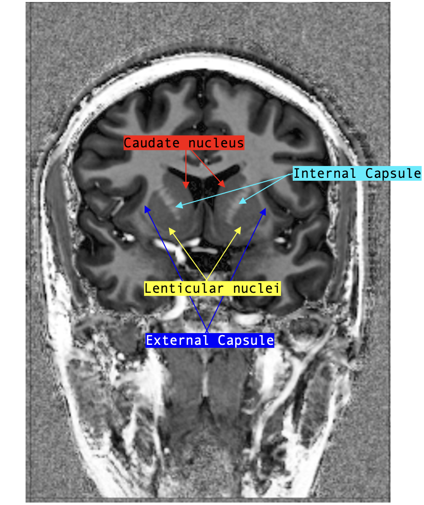
A few sections posterior to where the lenticular nuclei come into view, the thalami appear; the thalami are very large grey matter nuclei positioned right under the lateral ventricles (pushing the floor up).
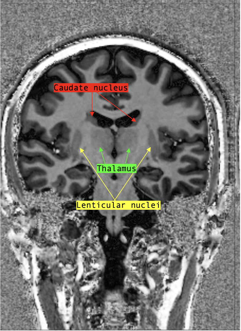
As the slices move more posterior, we see the caudate and lenticular nuclei taper and grow smaller while the thalami grow larger before eventually also tapering away.