All the images in this brick are in T1
Sagittal
The brainstem is composed of ascending and descending white matter tracts as well as multiple small grey-matter nuclei; as such, the overall intensity of the brainstem on MRI resembles more the intensity of white matter (ie. paler than grey matter), but with some heterogeneity.
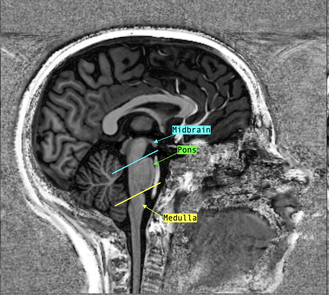
To start, find the mid-sagittal section; if it is exact, you should find the third ventricle but if you re slightly off-center, you will see a thalamus (a grey matter structure, therefore hyperintense than CSF) underneath the hyperintense curve of the fornix.
Whether you found the third ventricle or the thalamus, the midbrain begins just inferior to it. It may look like the beak of a bird in perfect mid-sagittal view.
Inferiorly, the midbrain is delimited by the pons which has a characteristic ventral bulge on sagittal.
The medulla is the most inferior portion of the brainstem; it begins superiorly where the pons ends (end of the “bulge”) and is delimited inferiorly by the foramen magnum, where it transitions into the spinal cord.
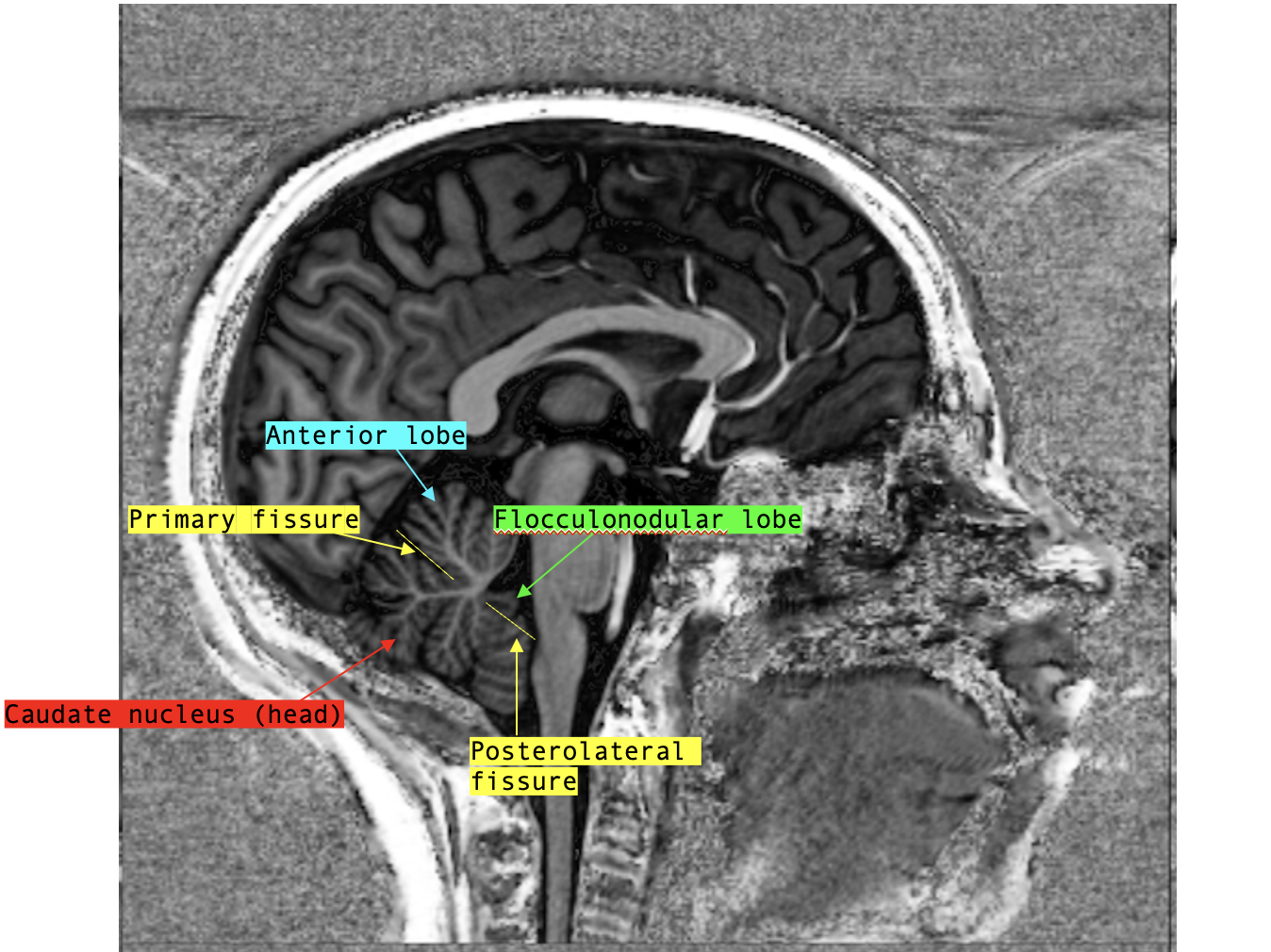
The lobes and fissures of the cerebellum can be well visualized on mid-sagittal as well.
Axial
On axial, it is important to recognize the level of the section by using nearby landmarks and the distinct cross-section shape of each part of the brainstem.
The cross-section of the midbrain has a distinct shape that resembles a Mickey Mouse head.
The “ears” of the midbrain correspond to the white matter tracts entering from the internal capsule!
To confirm that you are at the level of the midbrain, use the following landmarks:
- Only the inferior horns of the lateral ventricles should be visible
- The cerebral aqueduct should be visible
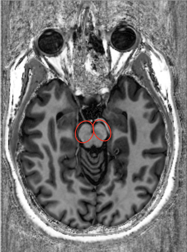
The cross-section shape of the pons evolves rostral to caudal, but there is always a little groove in the midline ventrally where the basilar artery can be seen running (see the red arrow in the image below).
That is why this is called the basilar groove!
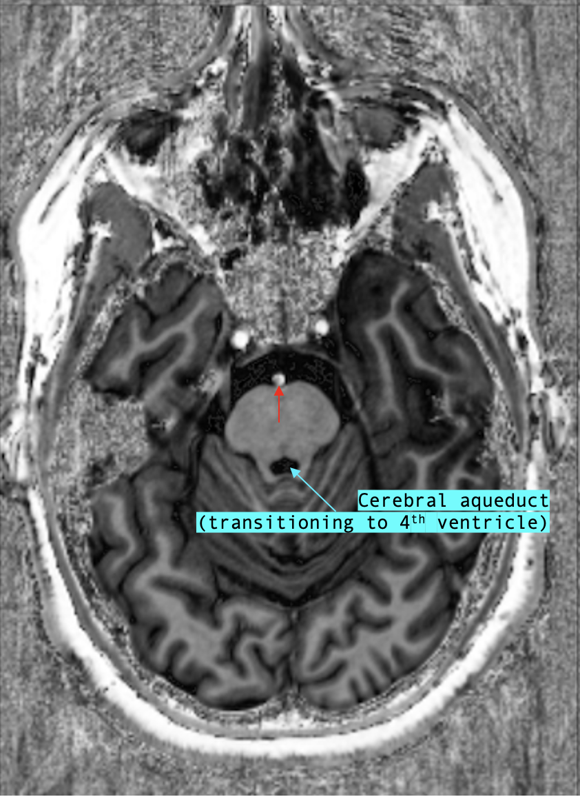
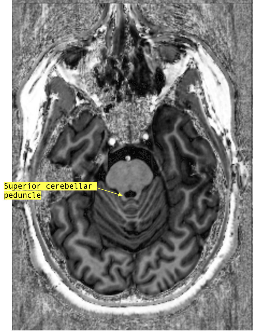
The cerebral aqueduct is still visible at the rostral pons, above the level of the superior cerebellar peduncle.
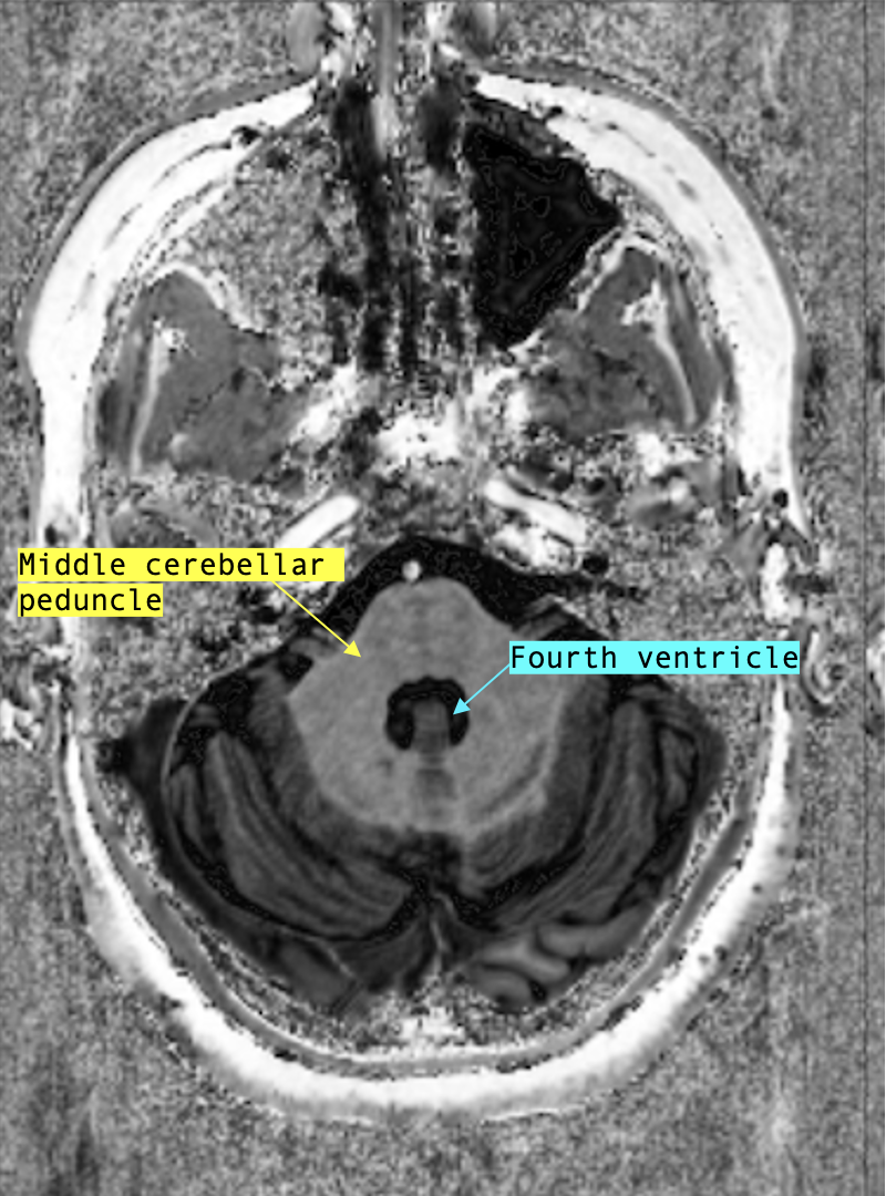
By the caudal pons it is the fourth ventricle that is visible instead. This is typically also the level where the middle cerebellar peduncles can be seen.
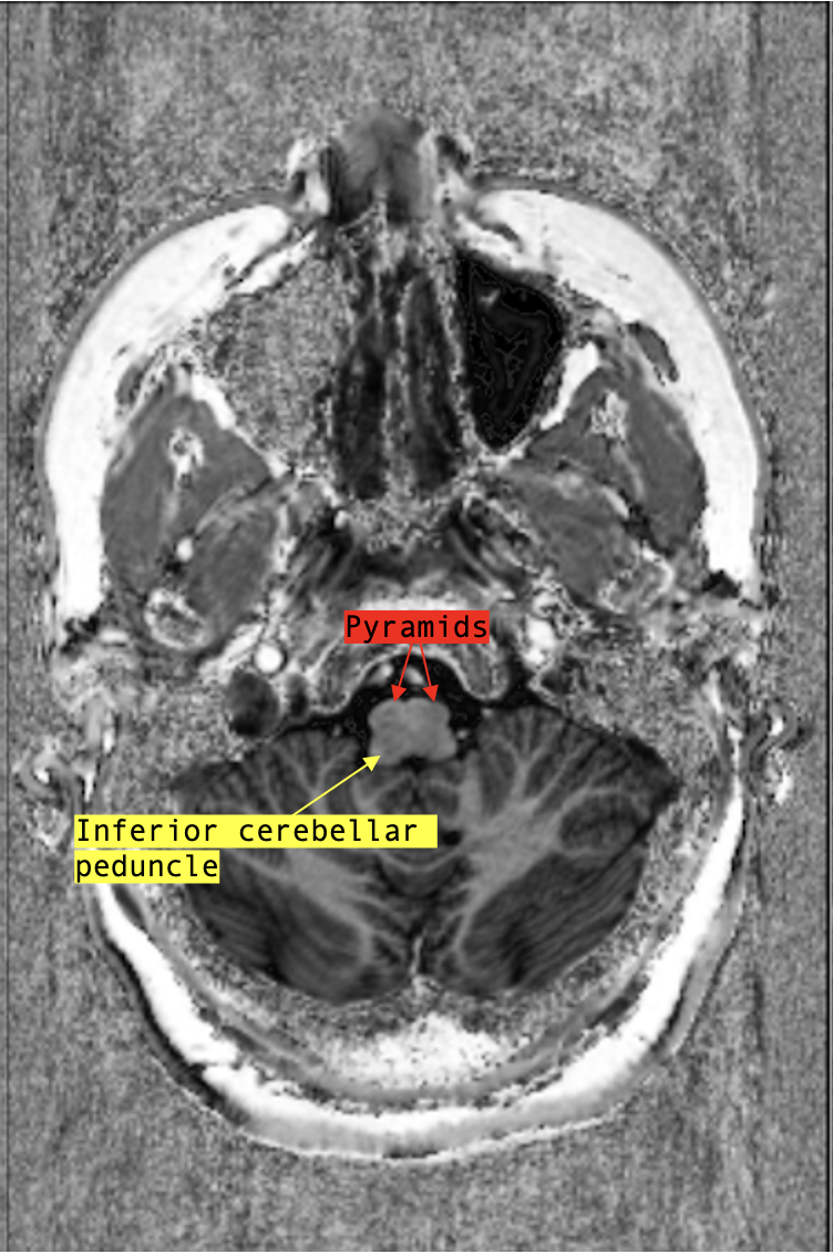
The cross-section of the medulla has two “pyramids” on its ventral aspect.
The inferior cerebellar peduncles can be seen connecting the medulla oblongata to the cerebellum at this level.