Ventricular System
The ventricular system consists of a series of ventricles and canals where cerebrospinal fluid (CSF) is produced and circulated.
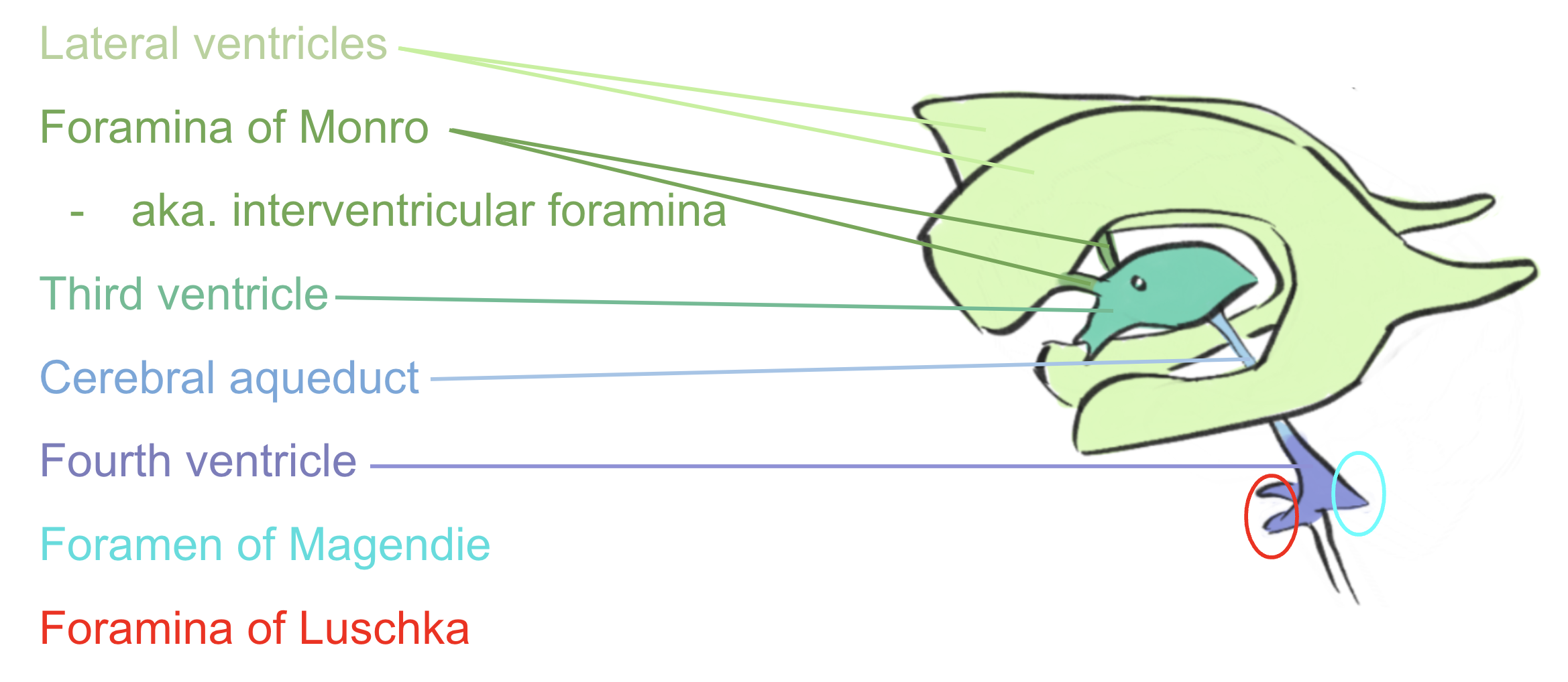
Figure 1: A 3D model of the ventricular system. In pale blue, we see illustrated the lateral ventricles. In turquoise, the bilateral foramen of Monroe can be seen connecting the lateral ventricles to the third ventricle, illustrated in pale yellow. The cerebral aqueduct is shown in red and connect the third ventricle to the fourth ventricle, shown in purple.
CSF is a sterile fluid that bathes the brain and the spinal cord. It provides buoyancy to the brain, alleviating pressure of gravity on its structures, as well as cushion from hits.
CSF also provides a chemically controlled environment for synaptic transmission reactions to occur. Recent findings even suggest that CSF flux may play a role in brain-wide waste clearance.
CSF is produced by the choroid plexus in the ventricles. The choroid plexus a complex network of capillaries lining each ventricles and produces CSF by filtering the circulating blood.
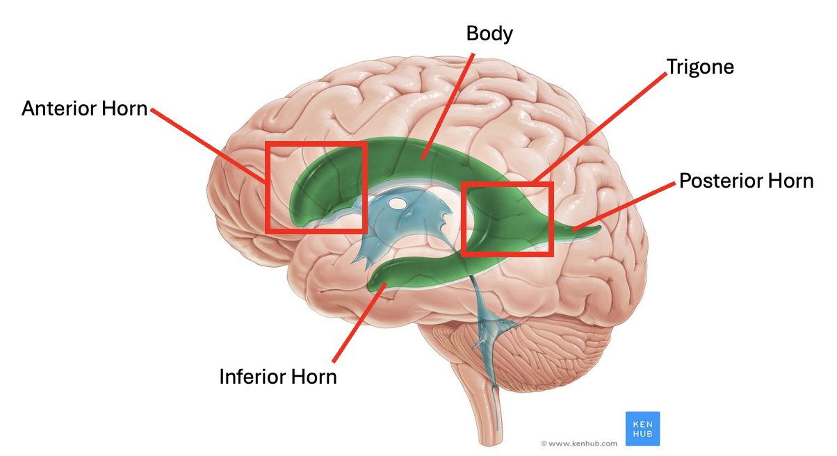
The largest two ventricles are the lateral ventricles. They are bilateral, CSF-filled structures that are located just under the corpus callosum of the cerebrum and connected by the interventricular foramen.
The lateral ventricles are divided into the anterior horns (that project into the frontal lobe), the body, the trigone, the posterior horns (that project into the occipital lobe) and the inferior horns (that project into the temporal lobe).
The lateral ventricles drain into the third ventricle via the foramina of Monro (one foramen per ventricle). It is located between the two thalami.
The anterior side of the third ventricle has two landmark protrusions: the supra-optic recess (right above the optic nerve) and the infundibular recess (right above the pituitary stalk).
The third ventricle then drains into the fourth ventricle via the cerebral aqueduct, a narrow space situated in the midline of the brainstem.
The fourth ventricle itself is located within the brainstem, at the junction of the pons and the medulla. It has a characteristic diamond-shape on axial cross-section.
The CSF in the fourth ventricle finally drains out via three caudal openings: two foramina of Lushka laterally, and one foramen of Magendie medially (hard to see on sagittal view).
The CSF exits into the subarachnoid space, the space between the pia and the arachnoid layers of the meninges. There, it is reabsorbed into the circulatory system by arachnoid granulations which protrude into the subarachnoid space and bring the CSF fluid into the dural venous sinuses.