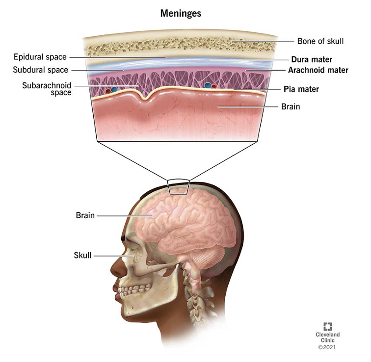
Meninges and meningeal folds
The meninges are three layers of connective tissue overlying the brain tissue (called the brain parenchyma). The meningeal layers are the dura , the arachnoid and the pia .
- Dura: the thickest and most superficial layer. The dura further separates into the periosteal and parietal layers; the periosteal dura is strongly adherent to the skull while the parietal layer slides against the arachnoid. The two layers stick tightly together, and only separate to form dural venous sinuses .
- Arachnoid: this layer sits between the dura and the pia. Granulations under this layer is responsible for absorbing CSF.
- Pia: this the thinnest and innermost layer. It generally cannot be separated from the brain tissue proper. Leptomeningeal vessels run along the surface of the pia.
The meninges form different folds that serve as important landmarks for neuroanatomy:
- Falx cerebri: separates the two cerebral hemispheres
- Tentorium cerebelli: horizontal fold that separates the cerebellum from the cerebrum
- Falx cerebelli: separates the two cerebellar hemispheres
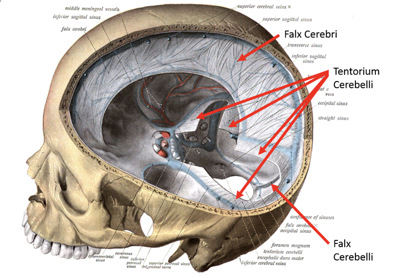
A trick: ‘falx’ usually refers to a vertical separating fold, whereas ‘tentorium’ separates structures horizontally.