Arterial Supply
Let’s start at the heart.
Oxygenated blood from the lungs enters the left heart which get pumped into the aorta with each systole. At this point, the blood splits into three different arteries: the left common carotid, the left subclavian and the brachiocephalic which itself quickly splits into the right subclavian and the right common carotid.
The two common carotid arteries (right and left) then travel up the neck and just below the jaw, they will branch into the external carotid and internal carotid arteries. The internal carotid arteries will then enter the skull through the carotid canal and to supply the brain with blood. However, given that the brain is the organ with the highest oxygen need per gram of tissue, evolution has created a second arterial supply.
From the subclavian arteries (right and left) we mentioned earlier arises the right and left vertebral arteries. Much like the carotids, the vertebrals also ascend the neck but by a more posterior route: threading through the transverse foramina of the cervical vertebrae, the vertebral arteries eventually enter the skull through the foramen magnum (through which the spinal cord passes).
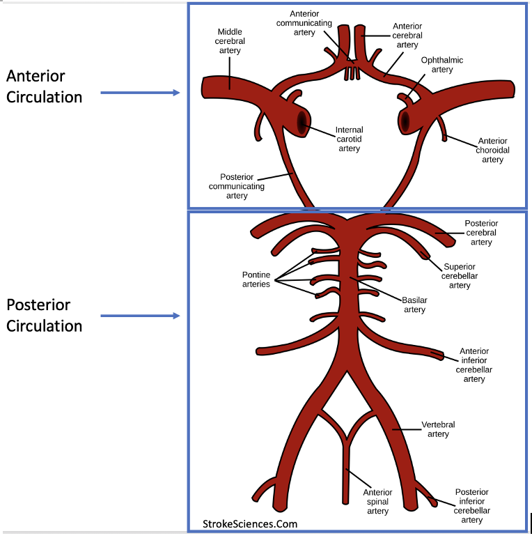
The two arterial supplies have been termed the anterior circulation (supplied by the internal carotid arteries and their branches) and the posterior circulation (supplied by the vertebral arteries and their branches).
The Circle of Willis
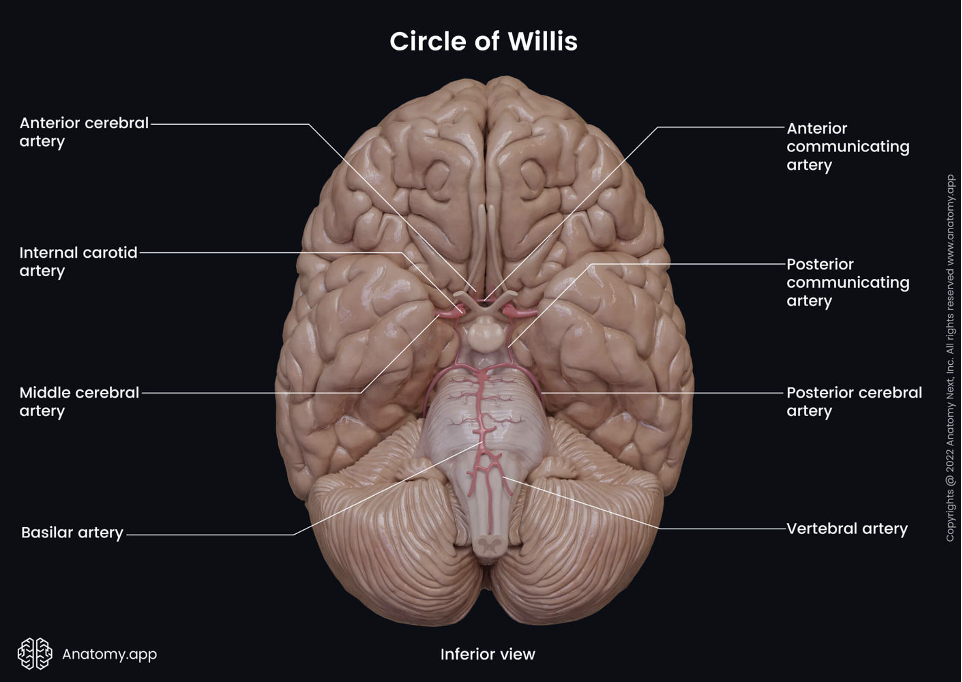
The anterior and posterior circulations connect at the Circle of Willis, a network of arteries that by its circular shape would ensure continued cerebral perfusion even if one of the two supplying circulations was compromised.
As seen on this model, the two posterior communicating arteries link the anterior and posterior circulations, allowing for collateral blood flow. It is useful to know the names and anatomical locations of all the arteries mentioned in this image since they, or their tributaries, are often involved in important stroke and hemorrhages.
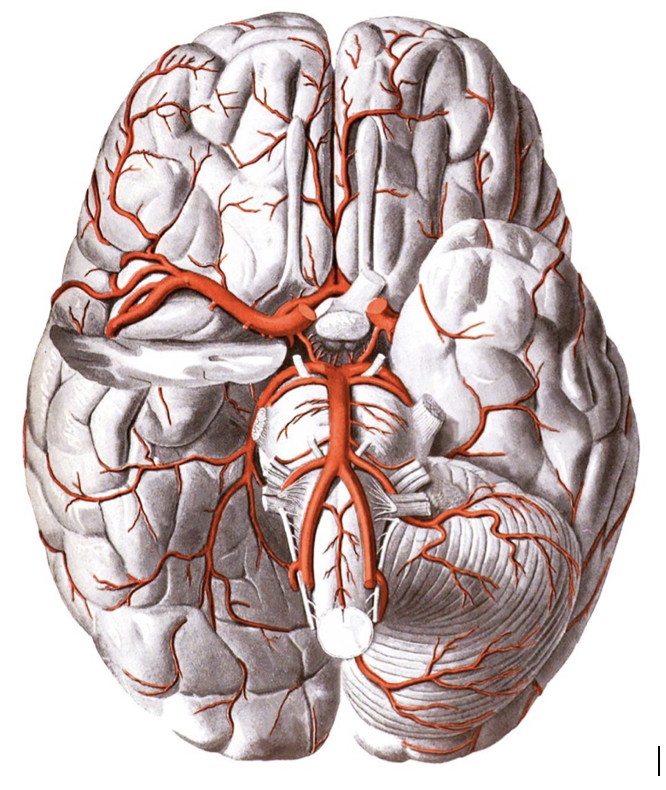
On a brain model:
Brainstem Perfusion
The brainstem is mainly perfused by the posterior circulation, like so:
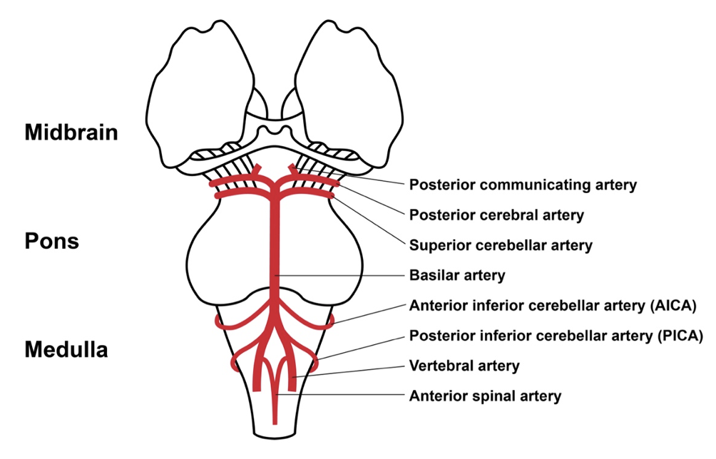
Each level of the brainstem will depend on different arteries for their blood supply; for instance, the upper portion of the Medulla is mainly perfused by the Basilar artery and the Anterior Inferior Cerebellar artery (AICA) but if you move slightly lower to the mid-Medulla, the AICA is replaced by the Posterior Inferior Cerebellar Artery (PICA) and the contribution from the Basilar artery gets smaller as you move down. For anyone who’s work pertains to stroke and/or hemorrhagic syndromes or those who want a solid foundation in neuroanatomy, we highly recommend checking out this page and to get familiar with the blood supply maps to the brainstem.
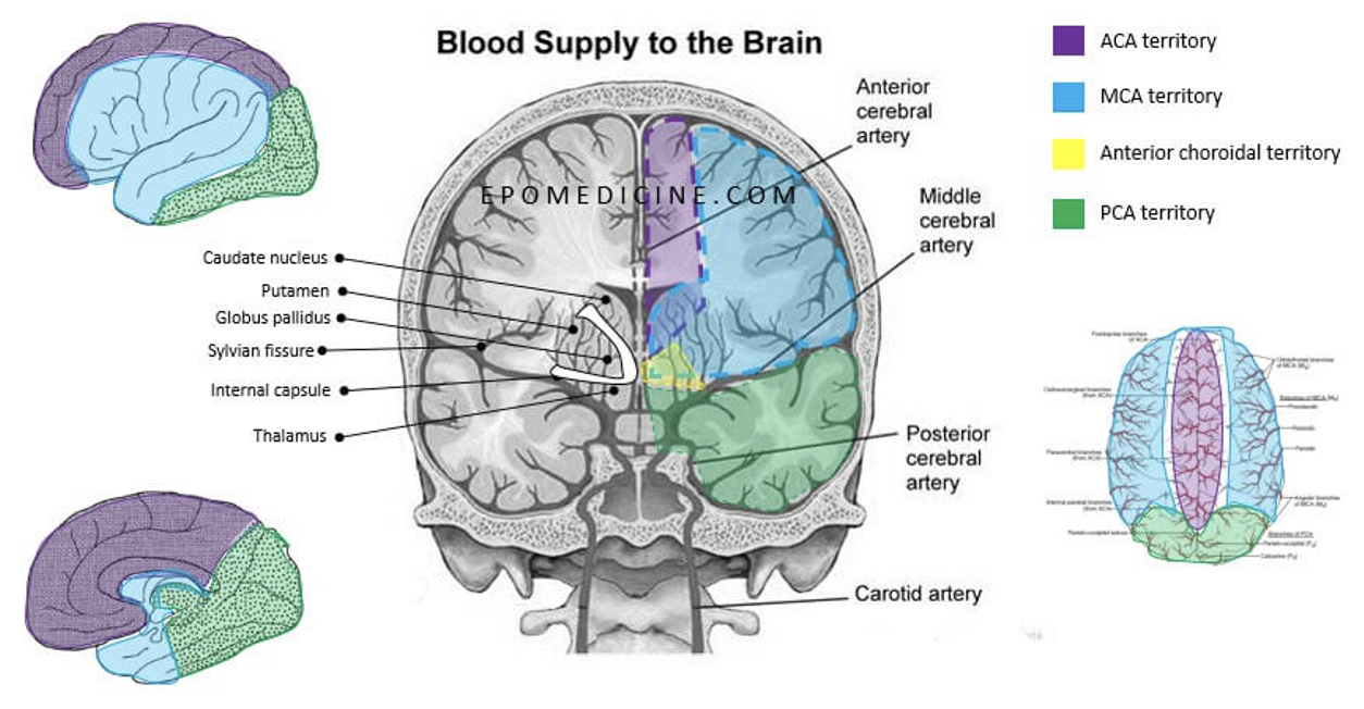
Cerebral Perfusion Territories
The cerebrum is mainly perfused by the Anterior, Middle and Posterior Cerebral arteries. In simple terms, the Anterior Cerebral artery (ACA) supplies the frontal lobe and the parts of the brain closest to the midline. The Middle Cerebral artery (MCA) supplies the lateral areas of both cerebral hemispheres, covering a vast area that includes most of the motor and sensory cortices. Branches from the MCA, often referred to as Lacunar or Penetrating arteries, travel deep inside the brain to feed the basal ganglia. Finally, the Posterior Cerebral artery (PCA) supplies the the posterior midline structures and the Occipital lobe.
Venous Drainage
Venous outflow doesn't follow arterial supply in the brain. Venous blood is drained in parallel by the superficial and deep systems; the superficial system is largely responsible for draining the cerebral cortex, whereas the deep venous system drains blood from the deep white matter and deep brain nuclei (eg. basal ganglia, thalamus). The deep venous system centers around the great cerebral vein (aka. the vein of Galen), which is an important landmark best appreciated on a mid-sagittal view.
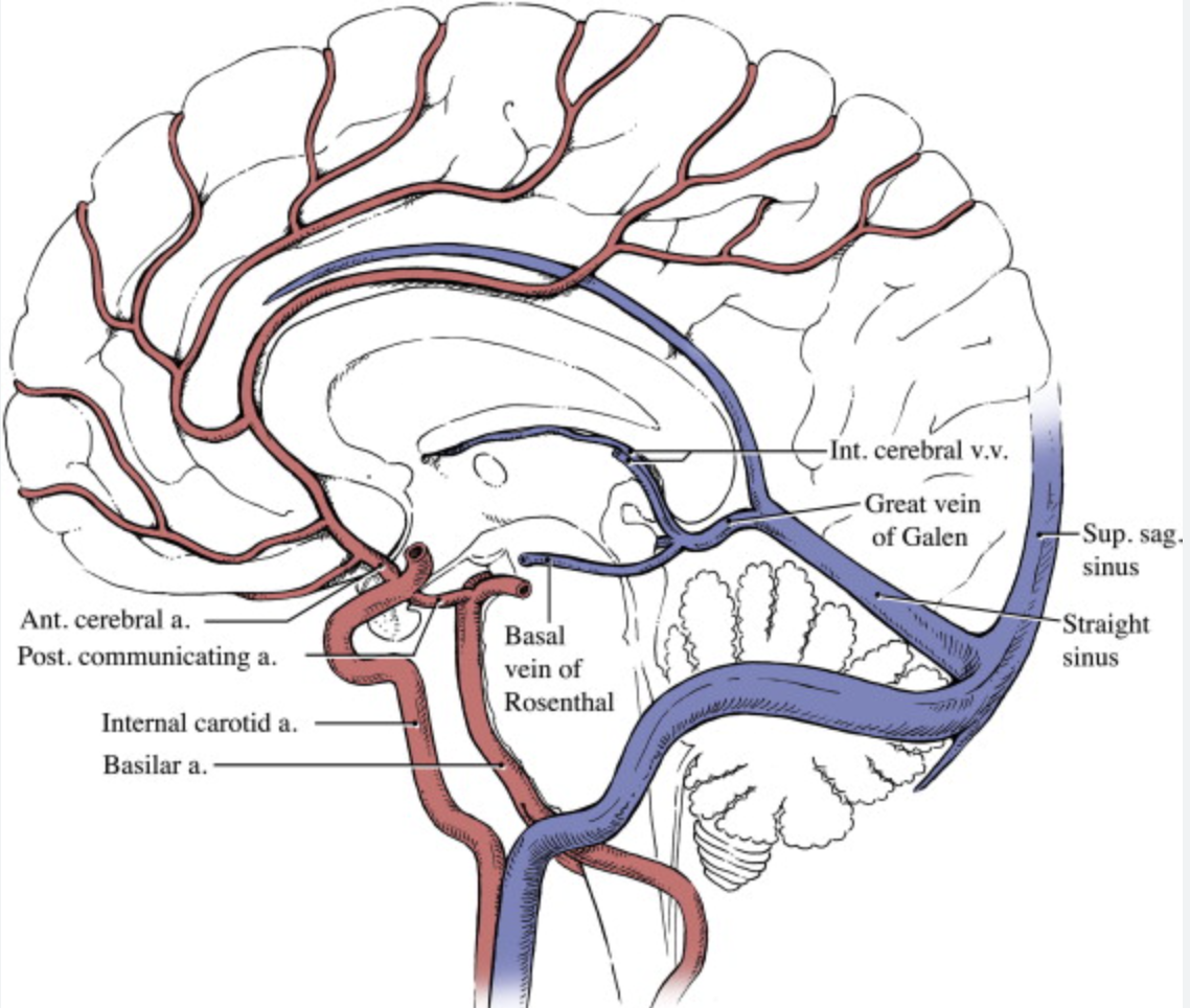
From these veins, venous blood is eventually drained into the dural venous sinuses.
These sinuses are pools of venous blood that are contained between the periosteal and meningeal layers of the dura mater- they receive all the venous drainage from the brain, but also get tributaries from the facial and scalp veins. The sinuses converge at the confluence of sinuses located in the occiput, before all eventually draining into the internal jugular vein.
Here is an image of the dural venous sinuses:
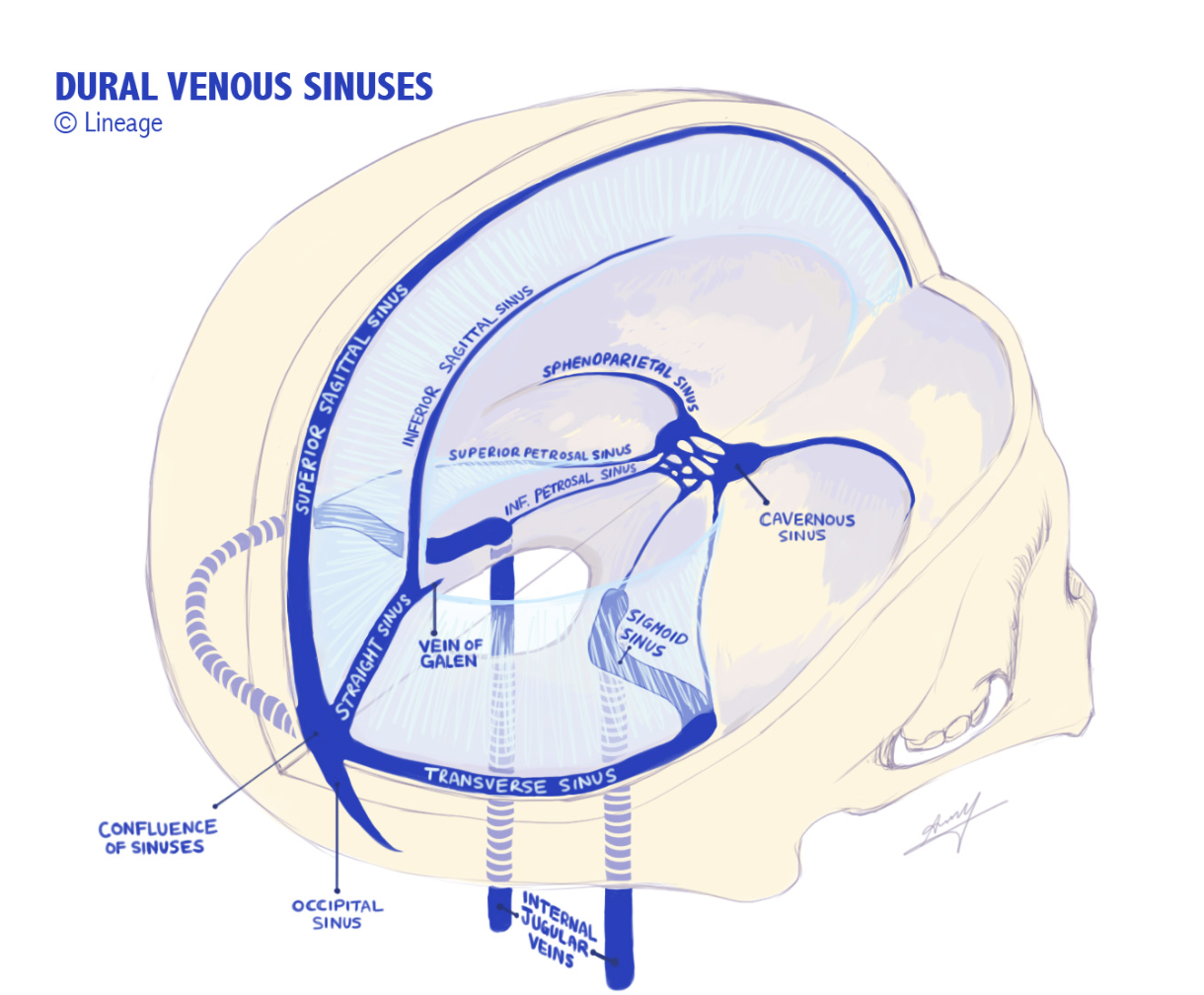
Notice how, from the confluence of sinuses, venous blood follows the transverse and sigmoid sinuses into the internal jugular veins.