In this chapter, we will discuss methods to find landmark gyri and sulci on imaging, focusing on sagittal views. These landmark gyri and sulci are less obvious on axial and coronal sections (particularly on static images), but we encourage you to practice looking for them in a scrollable DICOM image viewing software.
Sagittal
When approaching a sagittal section, an easy way to orient yourself is to find the midline.
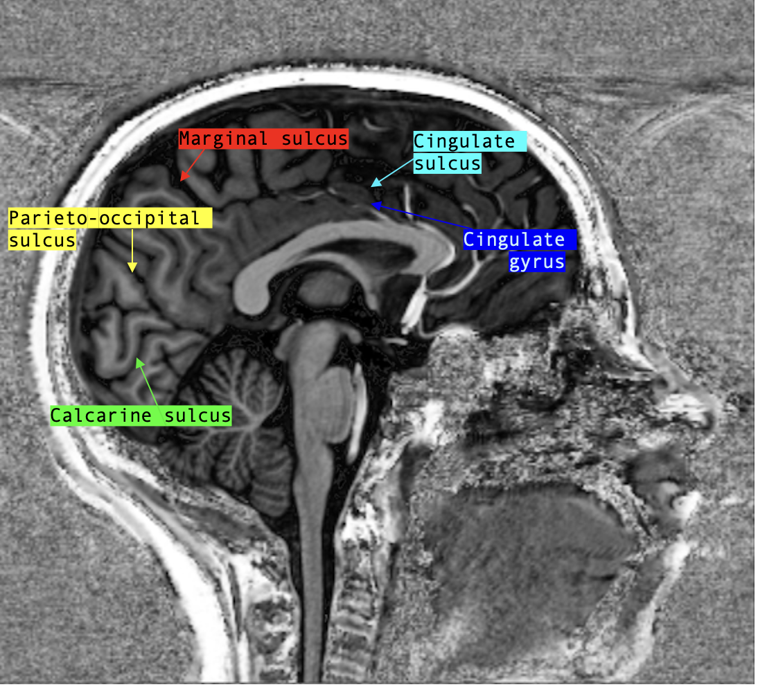
- Marginal sulcus:
- this sulcus is very easily found on mid-sagittal view and can serve as a landmark to identify many other structures so I like to start with it.
- Start by scrolling to the midline on the sagittal view.
- The marginal sulcus is found slightly posterior to the midline and is the only sulcus that descends directly from the cortex surface to the cingulate gyrus.
- The marginal sulcus can be found on axial view by starting from the surface of the cortex and scrolling down (caudally) all while paying attention to follow the sulci as you go down in order to identify which sulci persists until the cingulate.
- Parieto-occipital sulcus:
- found posterior to the marginal sulcus, the parieto-occipital sulcus originates from the surface of the cortex and curves deeper to end near the posterior aspect of the corpus callosum.
- This sulcus usually does not transect the cingulate gyrus , but rather appears to connect posteriorly with the cingulate sulcus.
- Calcarine sulcus:
- this sulcus is also most evident on mid-sagittal view.
- It travels from inferior aspect of the occipital cortex to the posterior aspect of the corpus callosum, where it joins with the parieto-occipital sulcus.
- What might be visually helpful is to remember that together, the calcarine sulcus and the parieto-occipital sulcus form a triangle (the cuneus).
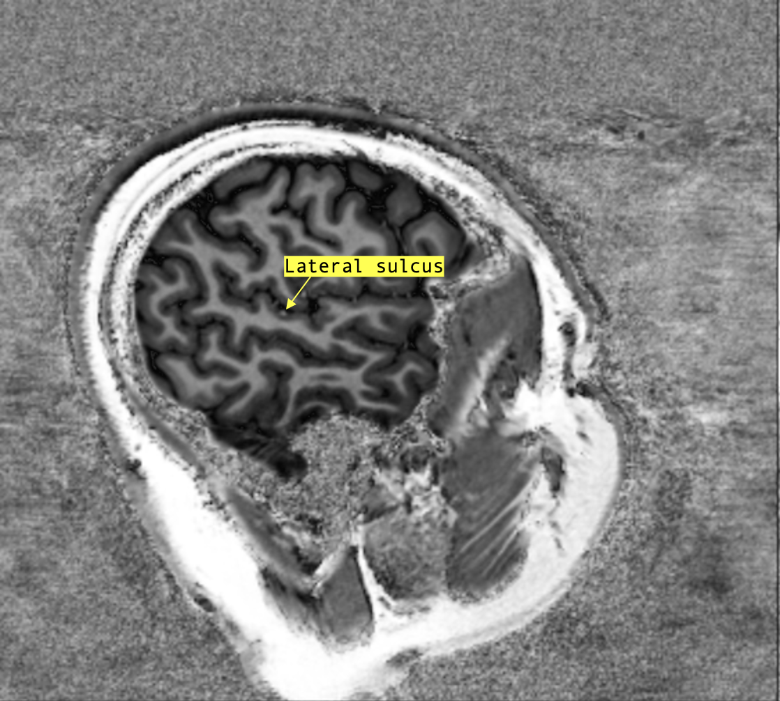
- Lateral sulcus
- can be appreciated on a superficial sagittal view (close to the surface of the brain).
- If the slice is superificial enough, you may also be able to appreciate the central sulcus .
From the marginal sulcus, find the sulcus immediately anterior: that is the central sulcus !
To confirm you have properly identified it, slowly scroll laterally while following the sulcus with your eyes (or finger!); the central sulcus joins with the lateral sulcus superficially without “breaking”.