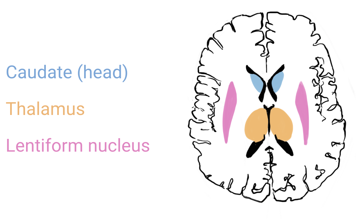
The basal ganglia are a collection of grey matter nuclei nestled deeply within the brain that are involved in many, complex brain functions. For instance, they act as a reward-predicting system that permits us to perform all kinds of daily tasks, from making the decisions about what to do next to fluidly coordinating that motion. They also play important roles in emotion, behavioural regulation, cognition, habit acquisition, procedural learning, and more! The basal ganglia has many components, the largest one being the corpus striatum , the subthalamic nucleus and the substantia nigra . The corpus striatum is then organized into the caudate nucleus and the lenticular nuclei (composed of the putamen and the globus pallidus).
Corpus striatum
The corpus striatum is structure of the basal ganglia that includes the following components:
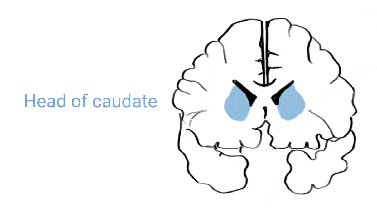
- Caudate nucleus - The caudate nucleus is a periventricular grey-matter structure divided into the head, the body and the tail. The caudate has a curved (C-like) shape in 3D that follows along the shape of the ventricles.
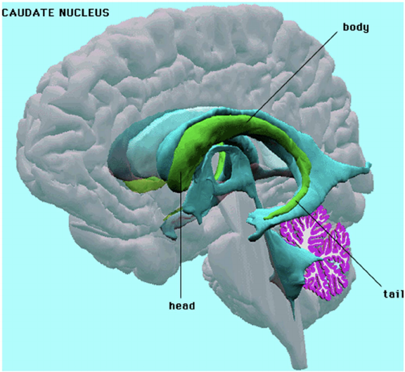
On coronal and axial sections, the head of the caudate (the biggest and most easily appreciated part) is found immediately lateral to the anterior horn of the lateral ventricle, nestled in the curve.
On sagittal sections, the caudate is not easily appreciated. It cannot be found at the midline since it is a structure nestled on the lateral aspect of each lateral ventricle.
- Lenticular nuclei - Bilateral grey-matter structures composed by the putamen and the globus pallidus.
Together, the putamen and globus pallidus form a wedge-like shape on both axial and coronal sections.
You can think of the lenticular nuclei together as a pizza-slice: the putamen is the crust and the globus pallidus is the part with the pall-atable toppings!
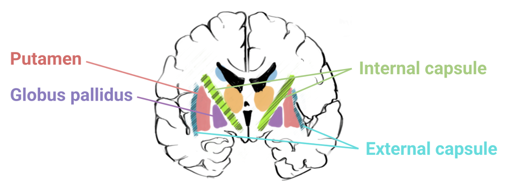
The putamen and globus pallidus are separated from the thalamus by the internal capsule (a highway of white matter tracts traveling between the cortex and the brainstem) and from the insular cortex by the external capsule.
Subthalamic nucleus
As per its name, the subthalamic nuclei are bilateral grey-matter structures located underneath each thalamus.
Substantia nigra
Located bilaterally underneath the subthalamic nuclei, neurons of the substantia nigra appear darker than surrounding tissues because of iron deposition. As such, the substantia nigra is fairly easy to appreciate with the naked eye on an anatomical slice.