Before we begin talking about basic neuroanatomy or neuroimaging, we must first introduce some terms that are frequently used to describe orientation or location in the brain.
Major Axes
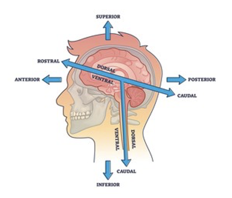
Often, in anatomy, locations of structures are described with "anterior," "posterior," "superior," and “inferior” in reference to the layman's terms of "front," "back," "up," and "down," respectively.
However, in neuroanatomy, you will also hear the words "rostral," "caudal," "dorsal," and "ventral." These terms originate from the embryological development of the central nervous system.
“Rostral” comes from the Latin word "rostrum," meaning beak or nose, and therefore refers to the very front of the head or face.
In contrast, “caudal” comes from the Latin word "cauda," which means tail; therefore, referring to the direction of the bum.
It is important to note that while the rostral-caudal axis may seem to overlap with the anterior-posterior axis, the rostral-caudal axis refers more to the directions of anatomical growth in embryology—the neural tube, precursor to the entire CNS, grew rostrally to form the brain and caudally to complete the spinal cord.
An easy way to visualize is to imagine a person on four-legs looking forward instead of standing. In this position, the anterior-posterior axis aligns with the rostral-caudal axis.
“Dorsal” and “ventral” are fairly descriptive and respectively mean “facing the back” or “facing the abdomen”. For any level below the brainstem, the dorsal-ventral axis generally map directly to anterior-posterior axis. However, since the brain curves anteriorly at the level of the cerebrum, the dorsal-ventral axis curves along with it so that “ventral” points more inferiorly and “dorsal” points more superiorly.
The image below summarizes these axes.
Relative positions
Many neuroanatomical terms are named in relation to another, nearby structure. Below are some examples with their explanations, though not all of these structures will be explored in this curriculum.
| Prefix | Meaning |
|---|---|
| Supra- | Above |
| Sub- | Below |
| Epi- | Upon/on |
| Peri- | Around/near |
Fiber directions
Afferent: of fibers, moving towards (when not referring to a particular structure, it generally means fiberes moving from the periphery to the CNS)
Efferent: of fibers, moving away (when not referring to a particular structure, it generally means fibers moving from the CNS to the periphery). Opposite of afferent.
Ascending: of fibers ascending from the peripheral nervous system (usually sensory fibers) to the CNS.
Descending: of fibers descending from the CNS to the periphery (usually motor or autonomic fibers). Opposite of ascending.